People sometimes ask me why anyone still draws plants and animals when every phone has a good camera. It is a fair question, and the answer explains what scientific illustration is.
A working definition
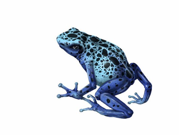
Scientific illustration is art made to communicate a scientific fact or idea accurately. The subject can be a species, an organ, a cell, a fossil, a layer of rock or a process such as metamorphosis. What makes it scientific is the purpose: the image exists to inform, and every choice in it, from the pose to the line weight, serves that purpose. A lovely drawing of a frog is not a scientific illustration if the toes are wrong.
That does not mean the work has to be dry. My own motto is "educational aesthetics." An image should be clear, accurate and interesting, and also beautiful. I believe the first step to taking care of our world is understanding it, and people are far more willing to study something that is a pleasure to look at.
Why a drawing can still beat a photograph
Photography is a wonderful tool, and I use photographs as references all the time. But there are jobs a camera does badly.
Clarity
A photograph records everything in front of the lens: the torn leaf, the shadow across the important part, the blurry background, the one specimen that happened to be available. An illustrator can choose. I can show a typical individual of a species instead of one particular animal, light it so the important structures read, and leave out whatever distracts. Field guides depend on this. Plates drawn in the same pose and light let a reader compare one species with the next in a way that photos taken in different conditions cannot.
Composites
A plant flowers in one season and sets seed in another. A drawing can show both on one page, along with enlarged details of the flower, the seed and the leaf surface. My plate of African rice (Oryza glaberrima) combines the whole plant with magnified views of a floret and the grains, information gathered from several references that no single photograph contains.
Cutaways and hidden structures
Some subjects cannot be photographed the way we need to see them: what lies under the skin, inside a seed or below the soil. For my life cycle of the chytrid fungus, I drew blocks of amphibian skin cut open to show the fungus growing inside. A microscope image shows one thin slice at a time. The illustration shows each stage whole, in context, with the frog it lives on at the center of the cycle.
Things no one can photograph
Extinct animals, ancient landscapes and past ways of life survive only as evidence: bones, pollen, pottery, the stains left by old posts in the soil. Reconstruction illustration turns that evidence into a picture people can understand. My graphite re-creation of daily life at the Kolb site, an archaeological site on the Pee Dee River in South Carolina, is that kind of image. No camera could have taken it.
The main branches
The field is wide, and most illustrators specialize in a few areas:
- Botanical illustration: plants, often as plates that combine the habit of the whole plant with the dissected details used for identification.
- Zoological illustration: animals from insects and fish to birds and mammals, including the species plates in field guides.
- Medical and anatomical illustration: human and animal anatomy, surgical procedures and patient education. This is a specialty with its own graduate programs.
- Paleontological and archaeological reconstruction: extinct life, past environments and human history, built from fossils and excavated evidence.
- Diagrams and process illustration: life cycles, food webs, cutaways and other explanatory figures for teaching.
My own work moves between several of these: plants like the stargazer lily and Euphorbia lactea, animals like the mimic octopus and kit foxes, the chytrid fungus life cycle, and the Kolb site reconstruction. You can see them in the gallery.
Accuracy and aesthetics
Scientific illustration is sometimes described as a tug of war between accuracy and art. I do not experience it that way. Accuracy comes first, always. If a composition would look better with a leaf turned, but the leaf does not grow that way, the leaf stays where it grows. Within the facts, though, there are hundreds of choices: the angle of view, which stage to show, how to arrange several views on a page, where to put the strongest contrast. Those choices are design, and good design is what makes a reader stop and look.
My Venus flytrap (Dionaea muscipula) is a good example. A single plant carries traps in different states at the same time, some open and some closed. Showing several of them together explains how the traps work, and the curves of the leaves also make the drawing graceful. The accurate choice and the attractive choice turned out to be the same choice.
Tools of the trade
Traditional media are still in daily use: graphite for soft tonal studies, pen and ink for line work that reproduces cleanly at small sizes, and watercolor or colored pencil for color plates. Digital painting has become standard for publication work because it makes corrections, labels and resizing much easier. Many illustrators work in both, sketching by hand and finishing digitally.
The less obvious tools matter just as much: a hand lens or microscope, calipers and a ruler, a good reference library, access to specimens, and a field journal. Observation and measurement come before drawing.
A very short history
Scientific illustration is as old as natural science. Early herbals used drawings so readers could recognize useful plants. Anatomists worked with artists to publish detailed atlases of the human body. Before photography, artists traveled with expeditions to record plants and animals that people at home had never seen, and painters such as Maria Sibylla Merian showed insects at every stage of their lives on the plants they fed on. Photography changed the field but did not replace it, for the reasons above. Today illustrators work for publishers, museums, research teams and educators.
How to get into scientific illustration
- Draw from life, constantly. Photographs flatten form, and drawing a real plant or animal teaches you how it is built. A field journal is the best training I know. I explain how I keep mine in How to Keep a Nature Field Journal.
- Study the science. At the Savannah College of Art and Design I majored in illustration and also took biology, anthropology and biomimicry. Knowing why a structure exists helps you draw it correctly.
- Learn the conventions. Scale bars, standard views, labels, plate layout and how images reproduce in print are all part of the craft.
- Build a portfolio of accurate work, and keep the references for each piece so you can show how you got there.
- Talk to scientists. Researchers, curators and nature center staff often welcome someone who can help explain their work, and their feedback will make your drawings better.
- Find your community. There are professional societies and certificate programs for natural science illustrators, and their members tend to be generous with advice.
If you need illustrations for a book, an exhibit or a classroom, take a look at my illustration services, especially scientific illustration for publishers, or get in touch.