The organism behind one of the worst wildlife diseases on record is too small to see, lives in the outer skin of frogs, and completes its whole life in a few days. That makes it hard to explain with words alone, which is exactly why I wanted to draw it.
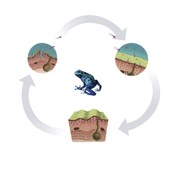
What the chytrid fungus is
Chytrids are an old and mostly modest group of fungi. Most live in water or damp soil, where they break down pollen, plant debris and other organic matter. They are unusual among fungi because they reproduce with spores that swim, each one driven by a single whip-like flagellum.
One chytrid broke the pattern. Batrachochytrium dendrobatidis, usually shortened to Bd, grows in the skin of living amphibians. It was described as a new species in 1999, after researchers working on sudden frog die-offs in Australia and Central America found a fungus in the skin of the dead animals. The name carries its history: batrachos is Greek for frog, chytra is an earthen pot (a nod to the pot-shaped spore case), and dendrobatidis refers to poison dart frogs. The organism used to define the species was isolated from a captive blue poison dart frog that died at the National Zoo in Washington, D.C. That is one reason the frog at the center of my diagram is a blue poison dart frog.
A second species, Batrachochytrium salamandrivorans, was described in 2013 and attacks mainly salamanders and newts. When people say "chytrid" in conversation about frogs, they nearly always mean Bd, and that is the one I drew.
The life cycle, stage by stage
The cycle is short and simple, which is part of why it spreads so well. In laboratory culture at around 22 °C it takes four to five days from spore to new spores, and researchers assume it runs on a similar schedule in skin.
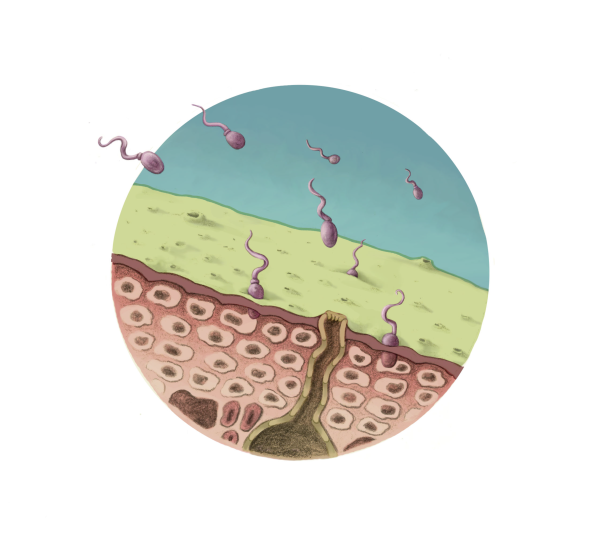
Stage 1: zoospores arrive
The infective stage is the zoospore, a single tiny cell with a flagellum. Zoospores swim through water or across a wet surface, but only over short distances, so infection usually comes from water, damp soil or direct contact with an infected animal. When a zoospore reaches amphibian skin it settles, absorbs its flagellum and builds a cell wall around itself. Biologists call this encysting.
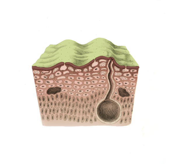
Stage 2: the sporangium grows inside the skin
The encysted spore sends a slender germ tube into the skin cells below. Bd lives only in the parts of the skin that contain keratin, the tough protein of the outermost cell layers. Inside a cell, the fungus swells into a round body called a zoosporangium, and its contents divide into many new zoospores. As the frog's skin cells mature and move outward, the sporangium is carried toward the surface with them, and a narrow discharge tube grows out to meet the open air or water.
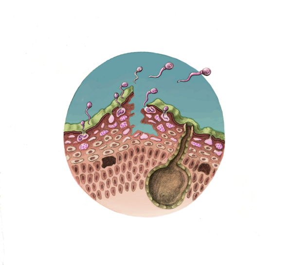
Stage 3: new zoospores escape
When the sporangium is ripe, the plug at the end of the discharge tube opens and the new zoospores swim out. Some leave the frog and find another host. Many simply settle on the same animal and start the cycle again, which is how a light infection can become a heavy one within a few weeks. The empty sporangium is shed with the old skin.
Why a skin infection can kill a frog
For us, skin is mostly a barrier. For a frog it is also a working organ. Amphibians drink and breathe partly through their skin, and they use it to move salts such as sodium and potassium in and out of the body. A heavy Bd infection thickens and disrupts the outer skin, and those exchanges start to fail. Research on infected frogs found that blood levels of key electrolytes fell sharply in sick animals, and the frogs died of cardiac arrest. Sick frogs often look lethargic, shed skin excessively and sit out in the open in a way a healthy frog would not.
Tadpoles, carriers and temperature
- Tadpoles: in most tadpoles the only keratin is in the mouthparts, so that is where the fungus lives. A tadpole can carry Bd without obvious harm and then sicken during metamorphosis, when keratin spreads across the new skin.
- Carriers: some species tolerate infection well. American bullfrogs and African clawed frogs are often named as hosts that can carry the fungus with few signs of disease, which lets them move it to new places.
- Temperature: Bd grows best in cool to moderate conditions and is slowed or killed by heat in culture. That is part of why outbreaks have hit many cool, high-elevation streams especially hard.
Where it came from and how it spread
Bd has been found on every continent where amphibians live. It has been linked to declines in hundreds of species, and to the disappearance of some species altogether. Genetic studies point to East Asia as the likely source of the most damaging lineage, and the global movement of amphibians for food, pets and laboratories probably carried it around the world. People can help spread it too, on wet boots, nets and gear moved from one pond to the next.
How I drew it
A life cycle diagram has to solve a few problems at once. Here is how I handled the main ones; I go into these principles in more detail on my page about life cycle diagrams for science education.
- Scale. A zoospore is microscopic and a frog fits in your hand. Putting the stages in round insets reads as "seen through a lens," so nobody thinks the spores are as big as the frog.
- Direction. The cycle runs clockwise from the top, and the arrows are big, soft and unlabeled. They only mean "this becomes that."
- Context. Each inset shows the same layers of skin, so the eye can follow the fungus moving from the surface down and back up again. The middle stage is a cut block rather than a circle, to show depth.
- The host. The frog in the middle ties the microscopic stages to an animal everyone can picture, and it reminds the viewer what is at stake.
- Color. Muted skin tones keep attention on the fungus, and the spores are the only purple in the image.
I drew each cutaway from micrographs and published descriptions, because there is no way to watch this happen with the naked eye. You can see all three stages at full size in the gallery.
What you can do
If you spend time around ponds and streams, simple habits help. Clean mud off boots and gear and let them dry completely before you visit another wetland. Avoid handling amphibians, and never release pet frogs, salamanders or aquarium water into the wild. I keep the same rules when I sketch in the field, as I explain in How to Keep a Nature Field Journal. If you find several dead or dying frogs in one place, report it to your state wildlife agency.
If you are teaching this topic, or explaining a disease, parasite or life cycle of your own, I would be glad to hear about the project. Get in touch.